 Aralens
Brain surgery neuronavigation system based on augmented reality
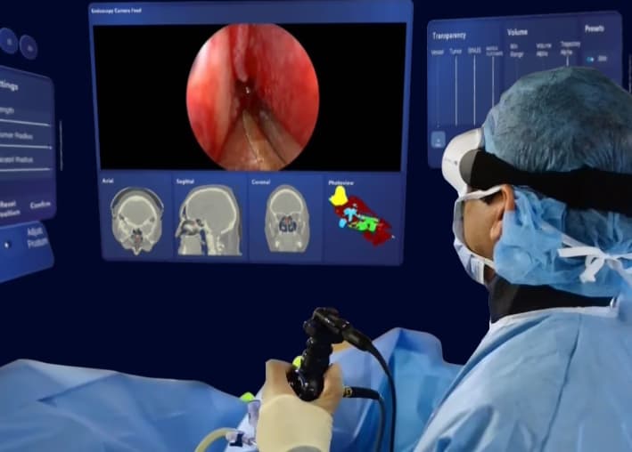
2D medical images do not always make the spatial relationships between vessels, tissue, and access routes immediately clear. Turning those images into practical guidance requires reliable spatial registration. Aralens, developed by Arassis within the YMOND ecosystem, addresses this need. It uses MRI and CT data for 3D processing and reconstruction, registration to patient anatomy, and augmented reality visualization to support surgical planning and guidance.

- Specialized department
- Arassis
- Scope of application
- Health and medicine
What exactly does this solution solve?
This solution is intended for neurosurgeons, biomedical engineering teams, teaching hospitals, and medical equipment procurement teams. It is designed for preoperative planning and spatial visualization of anatomical structures. Clinical claims require validation evidence. Implementation is tailored to each client's goals and operating conditions.
Start with the real problem: what does the end user need to do, which steps are difficult, what information or equipment is available, and how will success be measured? It uses MRI and CT data for 3D processing and reconstruction, registration to patient anatomy, and augmented reality visualization to support surgical planning and guidance. Defining these needs guides feature design and helps avoid unnecessary development costs.
Applications and use cases
- Review of MRI/CT and preoperative planning for brain surgery
- Co-registration of MRI and CT and volume reconstruction of the model
- Display model and segmented structures on patient anatomy
- Call saved path and entry point during operation
- Calibration and tracking of selected endoscope/instrument in AR view
Key capabilities and components
- Local or offline implementation can be considered in the technical scope of the project. Available modules and hardware models are specified in the proposal
- Aralens capabilities: workflow from MRI/CT co-registration to volume reconstruction and rendering, registration to real anatomy and calibration/tracking of instruments such as endoscopes
Core features, modules, and custom options are agreed during each project's needs assessment. The capabilities listed here describe the possible scope; they are not all necessarily included in a single package.
How the solution is experienced or implemented
Acquisition of MRI and CT images, with DICOM input based on project needs
Co-registration of two sets of images
Review/correction of segmentation and 3D model reconstruction with volume rendering
Check model in dashboard and plan/save entry point and route
Registration of patient model on actual anatomy
Calibration and tracking of selected instruments, such as endoscopes
Technology, requirements and project output
Technology and technical requirements
- Technologies and workflow: MRI/CT co-registration, three-dimensional reconstruction and volumetric rendering, registration on the real anatomy of the patient, calibration and tracking of surgical instruments including endoscopes
Customization and deliverables
- Configurable for selected data, anatomy, structures of interest, path/entry point, and surgical instrument. The user has raised potential development for ENT and orthopedics
- The paths of each discipline should be set with relevant tools, anatomy, workflow and validation. A customized project can be reviewed with pricing that is appropriate to the scope of work
- Changeable scopes and limitations are specified in the technical proposal
- The solution includes MRI/CT image streaming, co-registration, 3D reconstruction/volume rendering, registration to anatomy, planning, AR dashboard and selected tool tracking. One-time license, subscription, custom offer and pilot are some of the supply options. The breakdown of packages, equipment and the scope of each license must be specified in the proposal
Before starting, define the business or operational goal, an example scenario, budget and timing constraints, deployment environment, and technical decision-maker. These details help establish a realistic scope and delivery criteria.
Who is this solution for?
Purchasing and implementation may involve biomedical engineering teams and clinical decision-makers, depending on the intended use. It is designed for preoperative planning and spatial visualization of anatomical structures. Clinical claims require validation evidence. If the problem or access to the necessary data and content is unclear, start with a needs assessment or pilot.
Product image gallery
5 image


Related experience and projects
- Aralens; A platform developed in the field of neuronavigation and medical augmented reality
How to assess the solution's quality
Before purchasing or developing this solution, agree on the following three acceptance criteria in the technical review, alongside the required features:
- Technical and clinical validation for product claim scope
- Measurement of registration error, tracking and image data quality
- Review by a medical specialist and verification of regulatory documentation before clinical use
Frequently asked questions
How does augmented reality neuronavigation help planning?
3D reconstruction of medical images and spatial representation of the model can help the surgeon to examine the relationship of structures and design the access path. This application is not a substitute for clinical judgment
Can MRI and CT data be used?
The presented workflow includes MRI and CT data, co-registration, segmentation and volume representation. DICOM specifications and image quality should be checked before use
What role does surgical instrument tracking play?
Calibration and tracking of selected instruments such as endoscopes are reviewed in the technical scope of the project; The type of tracking system, accuracy and scope of application should be confirmed in the technical file
What documentation is required for clinical evaluation and purchase?
Evidence of validation, accuracy of registration, approved scope of use, necessary licenses, and documentation of professional liability should be reviewed before clinical use is claimed.
Why choose YMOND for this solution?
YMOND is a deep tech development group working across AI, computer vision, immersive XR experiences, and interactive hardware. Arassis is the specialist team behind this solution. Together, these capabilities allow the project team to address each client's challenge through coordinated software, content, and hardware design where needed.
How can I request a demo or a tailored proposal?
To explore neuronavigation for brain surgery for your organization, share your challenge, audience, operating environment, and expected outcomes with the YMOND team. Request a specialist consultation or start with the Vision Plus needs assessment to assess feasibility, discuss customization, and receive a technical proposal.
